
College of Dentistry at the University of Al-Qadisiyah Holds a Workshop on “The Importance of Radiography in Root Canal Treatment”
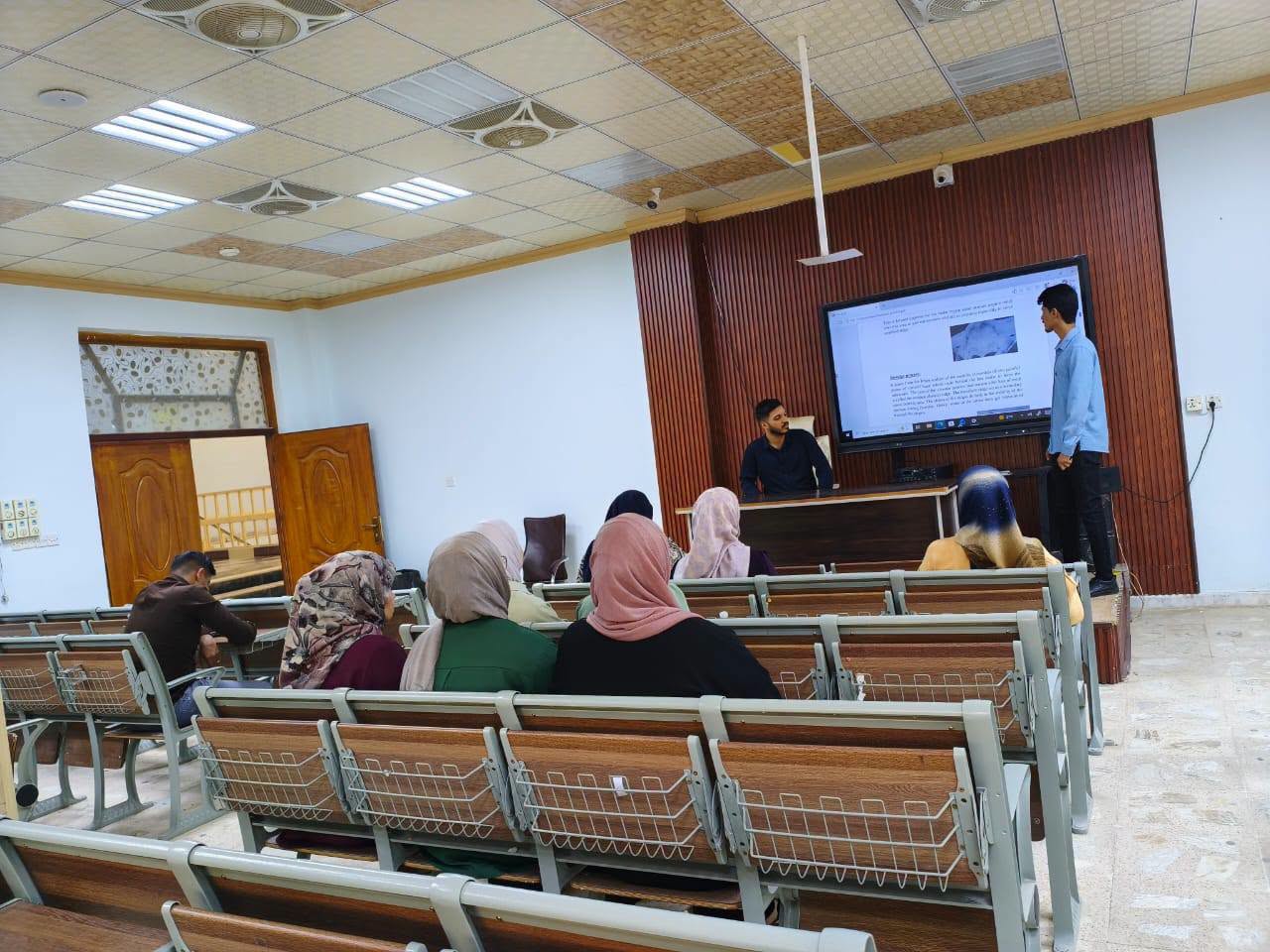
Under the patronage of the President of the University of Al-Qadisiyah, Prof. Dr. Rahi Kalf Mahdi Al-Yasiri, and the supervision of the Dean of the College of Dentistry, Prof. Dr. Ali Hassan Ali Murad, the Continuing Education Unit at the College of Dentistry organized a workshop entitled “The Importance of Radiography in Root Canal Treatment.”
The workshop was presented by the following trainee dentists:
- Ali Kamil
- Shakir Najm Abd
The workshop aimed to highlight the importance of dental radiography in diagnosing cases requiring root canal treatment, identifying the number, shape, and length of root canals before and during treatment, and accurately determining the working length to prevent instruments or filling materials from extending beyond the root apex.
It also addressed the role of radiographic imaging in evaluating root canal anatomy, detecting curvatures and anatomical variations, monitoring the different stages of root canal treatment, verifying the quality of canal cleaning and shaping, and assessing the final root canal filling in terms of length, density, and homogeneity.
The workshop recommended obtaining radiographic images before initiating root canal treatment to evaluate the condition of the tooth, its roots, and the surrounding tissues. Radiographs should also be taken during treatment when necessary, particularly to determine the working length and assess root canal morphology.
Furthermore, the workshop emphasized the importance of obtaining postoperative radiographs to evaluate the quality, length, and density of root canal fillings. It also recommended the use of digital radiography because of its rapid image acquisition, image enhancement capabilities, and ease of storage.
In complex cases where conventional radiography does not provide sufficient diagnostic information, such as the presence of additional canals or unusual root canal anatomy, cone-beam computed tomography (CBCT) may be considered when clinically justified.
The workshop also stressed the importance of minimizing patient exposure to ionizing radiation by applying appropriate radiation protection principles and limiting radiographic examinations to those that are clinically necessary.
Finally, participants were advised to compare radiographic images obtained before treatment, after treatment, and during follow-up visits to assess periapical healing and evaluate the success of root canal treatment.

لا تعليق